
If you’re like me, you’ve probably heard of or visited Philadelphia’s Mütter Museum. In 1939, the Kentucky Medical Journal of the Kentucky State Medical Association noted that in the Mütter was, and still is, a skeleton called the Kentucky Giant (sometimes referred to as the American Giant). The mounted skeleton is of an acromegalic giant and, at the time, was the largest and, “most spectacular that can be seen in America.”
Unknown Origins
The origin of the Kentucky Giant still remains a mystery. The body was secured in 1877 in an unknown location in Kentucky. It was largely believed that the body had been stolen from a cemetery. That same year, Prof. Joseph Leidy informed Prof. A. E. Foote that a body of a giant was offered for sale. However, no attempts at identification of the body were ever made. Dr. William Hunt arranged for the body to be transferred to Philadelphia and was then mounted by a Mr. R. H. Nash. The body was shipped to Philadelphia in a barrel of alcohol and sold for $500. By 1898, Leidy, Foote, Hunt, and Nash were dead which left little hope of ever identifying who the Kentucky Giant was.
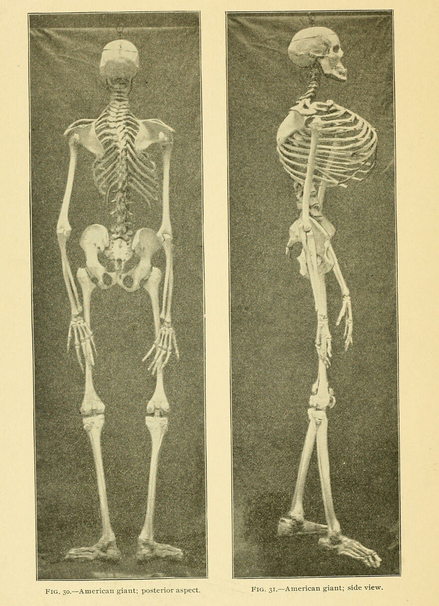
In 1898, Dr. Guy Hinsdale published an essay on acromegaly and in it was a very detailed description of the Kentucky Giant. It was believed, according to Dr. Hinsdale, that the Kentucky Giant was between 22 and 24 years of age upon his death as the bones seemed to have been fully developed by the time they were received by the Mütter Museum. “The epiphyseal junctions are plainly visible in all the long bones.” The spine underwent kyphoscoliosis, “which detracts considerably from he height which would otherwise exist.” As the skeleton was upon arrived, it measured seven feet and six inches.
No precise date of death was noted, though it’s possible that the young man died in 1876. Starting in 1939, there were calls for information seeking any information on a possible, “sickly and rheumatic giant, with malformed chest and twisted spine and a most ungainly frame, of unbelievably huge size, who lived a very obscure life in Kentucky.” The Kentucky Giant deserves a name and honor–not as a medical oddity, but as a human being.
Bones
Skull:

| Skull Measurement | Kentucky Giant |
| Cubic capacity, cc | 2320 |
| Length, glabella-occipital | 234 |
| internal | 195 |
| Height, basi-bregmatic | ………. |
| binaural over bregma | 351 |
| Vertical index | 78.3 |
| Breadth, maximum | 145 |
| minimum frontal | ………. |
| Cephalic Iindex | ………. |
| Horizontal circumference | 640 |
| Length of foramen magnum | 51 |
| Breadth of foramen magnum | 39 |
| Interzygomatic breadth | 147 |
| Intermalar breadth | 133 |
| Facial index | ………. |
| Orbital width | 52 |
| Orbital height | 42 |
| Orbital index | 80 |
| Palato-maxillary length | ………. |
| Palato-maxillary breadth | ………. |
| Palato-maxillary index | ………. |
| Pituitary Fossa | Kentucky Giant |
| Length | 27 |
| Depth | 17 |
| Breadth | 42 |
Face:
Dr. Hinsdale noted that the face was large but proportionate to the large cranium. Air sinuses were large and gave the great intermalar measurement, and the inferior maxilla, slightly prognathous, was massive. The mandible was a large bone measured as follows; the intercondyloid: 141, intergonial: 113, mentoalveolar: 40, width at angle: 33, width of ramus: 40, angle of ramus: 140 degrees.

| Vertical Depth of Face | Kentucky Giant |
| Length of face | 148 |
| Stature | 2285 |
| Face-stature index | 6.48 |
| Naso-alveolar length | 90 |
| Naso-alveolar stature index | 3.93 |
| Length of the face from nasion to chin compared with the size of the cranium. Circumference, 100 | 30 23.1 |
| Naso-alveolar length compared with size of the cranium. Circumference, 100 | 14.0 |
Vertebral Column:
According to Dr. Hinsdale, “the vertebrae are subject to conspicuous alteration, but they have been so mounted as to give a correct representation of the curves which existed in life.” Kyphoskoliosis was present, though not so much in the cervical vertebrae. Antero-posteriorly, there was a “sharp curve in the dorsal and lumbar region, with the convexity to the right. The kyphosis reaches its maximum at the ninth thoracic vertebrae, resulting in a compression and absorption of the body of that vertebrae.” Measurements of the anterior surface of the vertebral bodies are as follows: second thoracic: 30mm, third thoracic: 28mm, seventh thoracic: 25mm, ninth thoracic: 10mm, eleventh thoracic: 31mm, first lumbar: 36mm, second lumbar: 43mm, third lumbar: 45mm, fourth lumbar: 47mm, fifth lumbar: 50mm. The greatest width of the first thoracic vertebra was 97mm (3.75 inches). From the atlas to promontory of the sacrum: 820mm; the same points in a direct line were 750mm; from the atlas to the tip of the coccyx: 1030mm.
The lumbar vertebrae were massive with the sacrum being composed of four vertebrae instead of five. The width being 16cm and the length being 13.2cm. The coccyx consisted of three vertebrae instead of four. The ribs were long and narrow and relatively straight. The seventh and eighth ribs measured 45cm along the under border and 43.5cm in length; the sixth rib on the left side on the outer side was 43.5cm long; the seventh, measured along the under side, was 43.8cm in length.
The thorax was large but narrow in proportion to its depth. The girth was 109.7cm (43.25 inches); antero-posterior diameter was 43cm; lateral diameter was 28cm. It was theorized that in individuals with acromegaly that the thorax was relatively narrow.
Pelvis:
The pelvis was large but proportionate to its overall size. Bones were thick at their borders and bore marks of periosteal inflammation. The iliac crests and above the acetabula were arthritic. “The conformation is of the rachitic type, judging from the increase of the measurement between the anterior superior spines and the crests. The cavity of the pelvis is considerably encroached upon by the convexities which mark the position of the acetabula, particularly that of the left side. The acetabula are very deep and separated by thin bone from the pelvic cavity. The bodies of the iliac bones are exceedingly thin.”
| Pelvic Measurements | Kentucky Giant |
| Between the anterior superior spines | 33.5cm |
| Between the crests | 33.5 |
| Between the middle points of the ischii | 15.5 |
| Antero-posterior diameter of pelvic inlet | 14 |
| Left anterior oblique diameter of pelvic inlet | 17.5 |
| Left anterior oblique diameter of pelvic inlet | 17.7 |
| Depth of pelvic cavity | ………. |
| Height of pelvis | ………. |
Upper Extremities:
The clavicles were 212mm in length; the scapulae were, in length, acromion to angle, R. 268mm, L. 268mm and breadth, R. 144, L. 150. Length of the right humerus was 47.5cm with a circular perforation in the olecranon fossa that was 16mm in diameter. The right ulna measured 37.8cm, the left, 37.5cm; right radius measured 354, the left measured 360; the length of the hand from the scaphoid to the tip of the the middle finger was 25cm.
Lower Extremities:
The femora were slender but appropriately proportioned given the size, they were symmetric and the shafts were well-formed and unduly curved. The right femur measured 65.5cm, the left measured 66.6cm. The shafts were 84 and 85mm in circumference for the right and left sides respectively, and 20mm in diameter. Accordingly, the shafts were no thicker than compared to average-size individuals. The hips joints were “markedly arthritic,” and the femoral heads were misshapen and the necks deformed. Rather than the necks being circular in shape, they were semilunar, short and approached a right angle in relation to the shaft. Circumference and greatest diameter of the necks are measured as follows: right, 13.5, left, 13. Greatest diameter of right was 5.7 with greatest diameter of left being 5.5. Condyles were well formed. “The compact tissue is very thin at the extremities, barely covering the cancellous structure.” The fibulae curved with their convexity backward, so that the perpendicular to the chord of the arc is 47mm. The middle portion of the shaft in both cases lies entirely posterior to the tibiae”
Contributed by Shawn Logan | contact@kyhi.org
⁘ Works Cited ⁘
- Acromegaly: an essay to which was awarded the Boylston Prize of Harvard University for the year 1898 / by Guy Hinsdale. (2025). Wellcome Collection.
- Kentucky Medical Journal, Vol. 37, February 1939.
Important note:
If you would like to use any information on this website (including text, bios, photos and any other information) we encourage you to contact us. We do not own all of the materials on this website/blog. Many of these materials are courtesy of other sources and the original copyright holders retain all applicable rights under the law. Please remember that information contained on this site, authored/owned by KHI, is provided under a Creative Commons Attribution-NonCommercial-NoDerivatives 4.0 International License.
Photographs, text, illustrations and all other media not authored by KHI belong to their respective authors/owners/copyright holders and are used here for educational purposes only under Title 17 U.S. Code § 107.

